
Retina Treatment

The retina is a light-sensitive layer of tissue located at the back of the eye. It plays an essential role in vision by converting light into signals that are sent to the brain through the optic nerve. Any damage to the retina can affect central or peripheral vision and may lead to serious vision problems if left untreated.
At Shree Hanumant Vision Care (SHVC), Varanasi, we focus on eye examinations and the evaluation of retinal conditions to help identify vision-related problems. Early detection, appropriate diagnosis and timely medical care are important for maintaining long-term retinal health.
What Are Retinal Diseases?
Retinal diseases are eye conditions that affect the retina and its ability to process visual information. These conditions may develop due to ageing, diabetes, changes in retinal blood vessels, eye injuries or other underlying health problems.
Some retinal conditions develop gradually without noticeable symptoms, while others may cause sudden changes in vision. Regular eye examinations can help detect retinal abnormalities at an early stage.
Common Types of Retinal Diseases
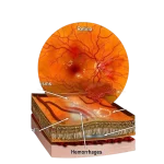
Diabetic Retinopathy
A condition in which high blood sugar damages the small blood vessels of the retina, potentially causing swelling, bleeding and vision changes.
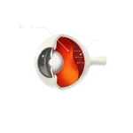
Retinal Detachment
Occurs when the retina separates from its normal position at the back of the eye. It requires urgent medical attention.
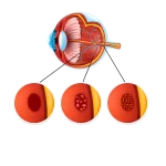
Age-Related Macular Degeneration (AMD)
A condition affecting the macula, the central part of the retina responsible for detailed vision.
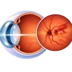
Retinal Vein Occlusion
A blockage in a retinal vein that can cause blood and fluid to leak into the retina, affecting vision.
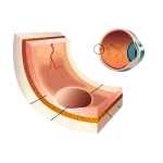
Macular Hole
A small opening in the macula that may cause blurred or distorted central vision.
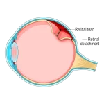
Retinal Tear
A break or small opening in the retina that may progress to retinal detachment if not treated appropriately.
Causes and Risk Factors of Retinal Diseases
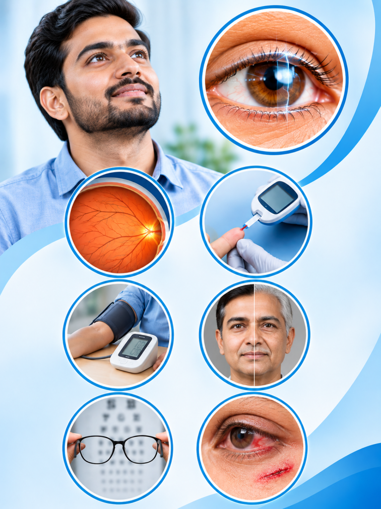
Retinal diseases can develop because of factors that affect the blood supply, structure or health of the retina. Some conditions are linked to age or genetics. Others are closely connected to lifestyle conditions such as diabetes and high blood pressure. Knowing your risk factors helps you take timely preventive steps.
Common causes and risk factors include
Diabetes: Long-term or poorly controlled blood sugar can damage the retinal blood vessels and lead to diabetic retinopathy.
High Blood Pressure: Uncontrolled hypertension can strain and damage the delicate vessels of the retina.
Increasing Age: The risk of macular degeneration, macular holes and retinal vein occlusion rises with age, especially after 50.
High Myopia (Short-sightedness): People with high minus power have a thinner retina, which may increase the risk of retinal tears and detachment.
Eye Injury: A blow or injury to the eye can cause bleeding, tears or detachment of the retina.
Family History: Some retinal conditions, such as retinitis pigmentosa and certain types of macular degeneration, can run in families.
Smoking: Smoking is a known risk factor for macular degeneration and can harm overall blood vessel health.
Retina Treatment Options
How are retinal diseases treated?
Treatment depends on the type, stage and cause of the retinal condition. After a detailed examination, the eye specialist may recommend
- Regular monitoring and follow-up for early-stage conditions
- Better control of blood sugar and blood pressure, together with your physician
- Medicines or eye drops to manage swelling and inflammation
- Intravitreal injections (such as anti-VEGF) for certain macular and retinal conditions
- Laser treatment to seal leaking blood vessels or treat retinal tears
- Surgical treatment, such as vitrectomy, for advanced or complex cases
- Low-vision guidance and lifestyle advice to protect remaining vision
Treatment results vary from patient to patient. Early diagnosis usually gives more options and a better chance of protecting vision, which is why timely consultation with an eye specialist matters.
How Are Retinal Diseases Diagnosed?
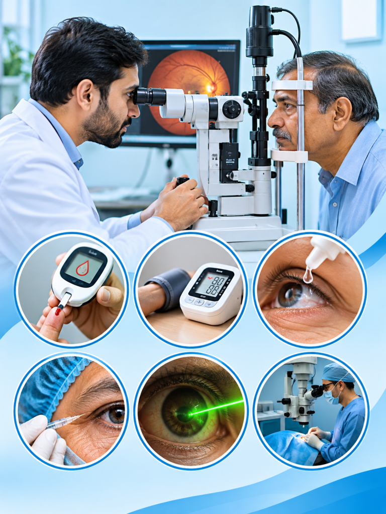
A detailed eye examination is important for finding the type and severity of a retinal condition. Depending on your symptoms and medical history, an ophthalmologist may recommend different diagnostic tests.
- Dilated Fundus Examination: Eye drops are used to widen (dilate) the pupil, so the doctor can examine the retina, macula and optic nerve in detail.
- Optical Coherence Tomography (OCT): This non-contact scan creates detailed cross-section images of the retina. It helps detect swelling, fluid or thinning in the macula.
- Fundus Photography: High-resolution photographs of the retina are taken to record its condition and track changes over time.
- Fundus Fluorescein Angiography (FFA): A special dye is injected into a vein in the arm, and photographs show how blood flows through the retinal vessels. This helps identify leakage or blockages.
- B-Scan Ultrasonography: This ultrasound test helps assess the retina when the view is blocked by bleeding or a dense cataract. It is useful for detecting retinal detachment. The tests recommended depend on each patient’s condition and examination findings.